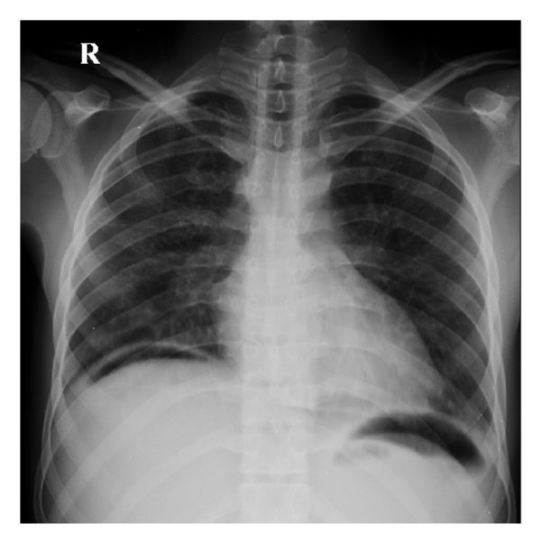
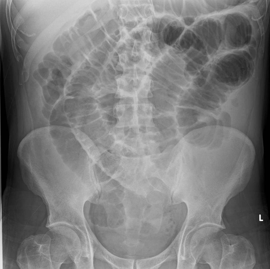
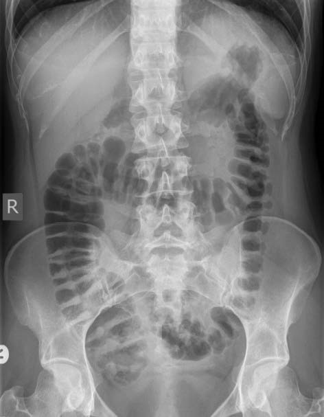
ABDOMINAL X-RAY (AXR) / MASTERCLASS, SECTIONS / By TopicsNotesBowel Diameter:MediaReferences Topics Ryles Tube – indications, testsNJ Tube AOD – Commonest 2 causes AXR: BowelBonesCalcification(Stones) The upper limits for the normal diameter 3,6,9 rule (SBO >3cm LBO >6cm Caecum >9cm) LBO & Ileo-Caecal Valve Significance Notes Projection of the abdominal X-ray.Typical projections of an abdominal X-ray include:Anterior-posterior (AP) supineAnterior-posterior (AP) erectBBC approach:Bowel and other organs: small bowel, large bowel, lungs, liver, gallbladder, stomach, psoas muscles, kidneys, spleen and bladder.Bones: ribs, lumbar vertebrae, sacrum, coccyx, pelvis and proximal femurs.Calcification and artefact (e.g. renal stones)Bowel Diameter:The upper limits for the normal diameter of different bowel segments are as follows:Small bowel: 3cmColon: 6 cmCaecum: 9 cmThis is often referred to as the ‘3/6/9 rule’. Media Air Under Diaphragm Faecal Lading SBO Gaseous Large Bowel Coffee bean isolated on white background References Link Link Link Link