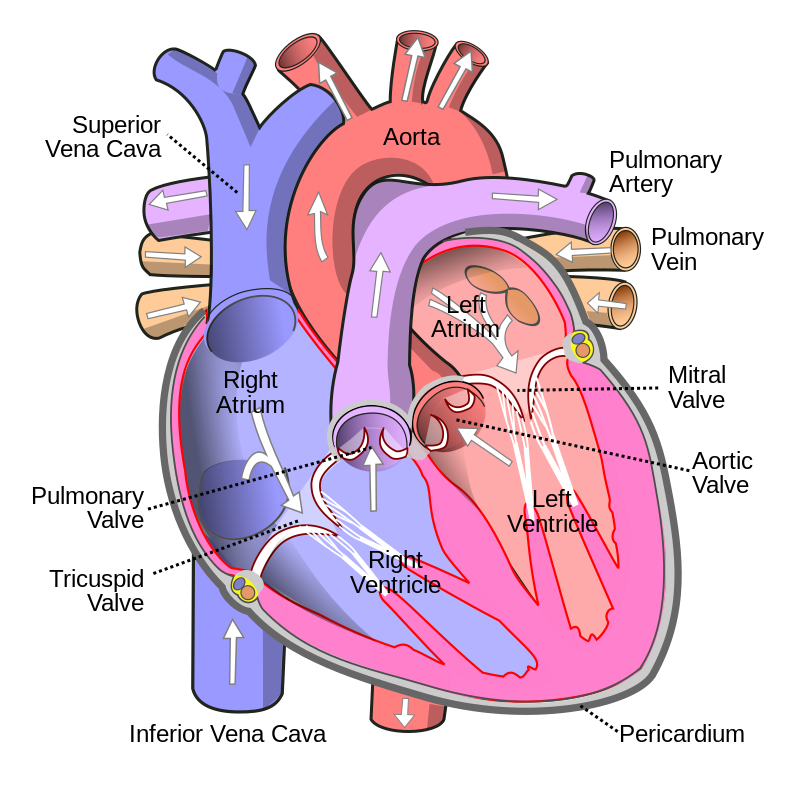
- Blood Supply Heart
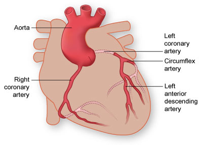
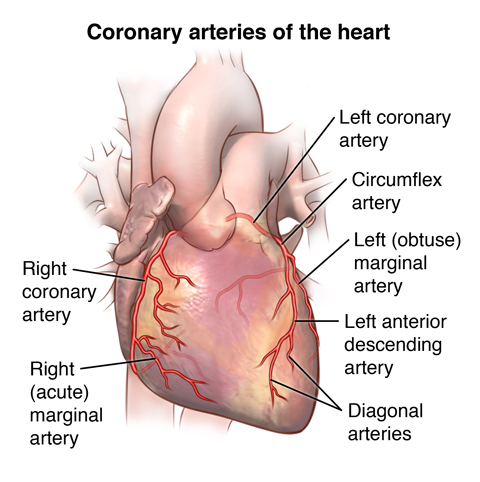
What are the different coronary arteries? The 2 main coronary arteries are the left main and right coronary arteries.
-
Left main coronary artery (LMCA). The left main coronary artery supplies blood to the left side of the heart muscle (the left ventricle and left atrium). The left main coronary divides into branches:
The left anterior descending artery LAD branches off the left coronary artery and supplies blood to the front of the left side of the heart.
The circumflex artery branches off the left coronary artery and encircles the heart muscle. This artery supplies blood to the outer side and back of the heart. -
Right coronary artery (RCA). The right coronary artery supplies blood to the right ventricle, the right atrium, and the SA (sinoatrial) and AV (atrioventricular) nodes, which regulate the heart rhythm. The right coronary artery divides into smaller branches, including the right posterior descending artery and the acute marginal artery. Together with the left anterior descending artery, the right coronary artery helps supply blood to the middle or septum of the heart.
Smaller branches of the coronary arteries include: obtuse marginal (OM), septal perforator (SP), and diagonals.
- Clot/Thrombus/Embolus
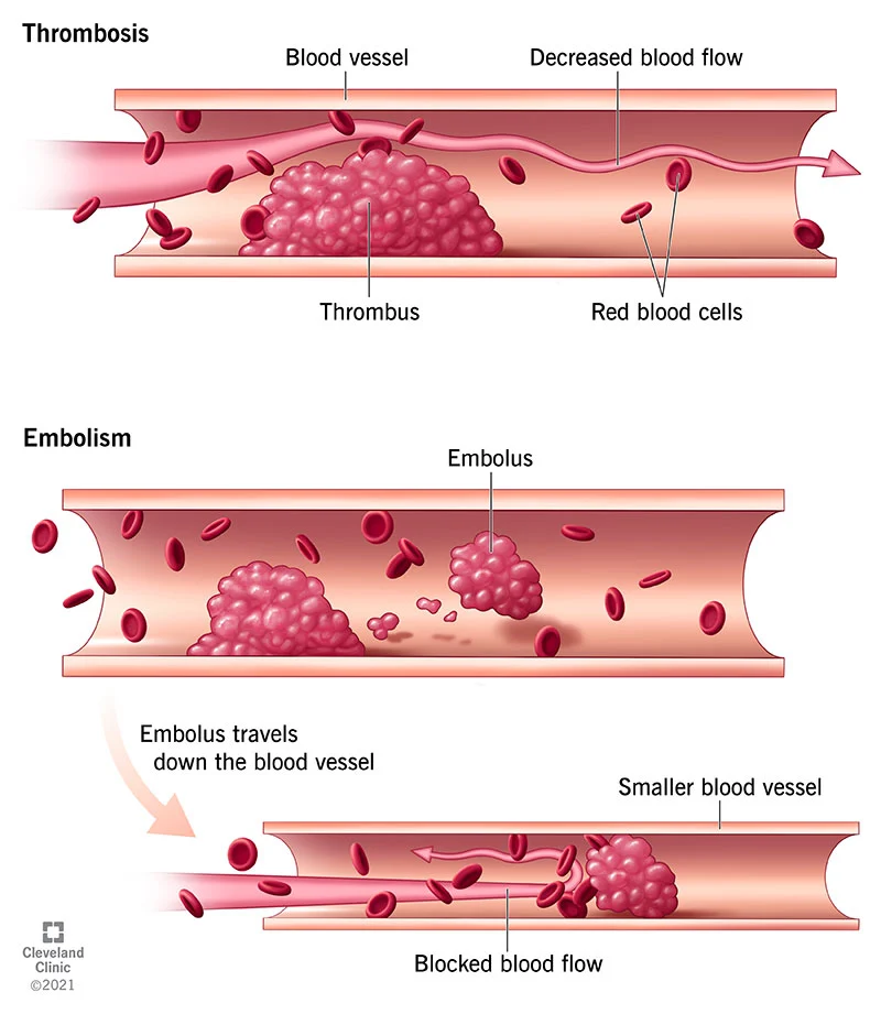
Embolus: An embolus is a particle or mass that flows through the bloodstream. A thrombus is a blood clot in a blood vessel. If a thrombus breaks off, it can become an embolus. Link
Plaque: Plaque is made up of deposits of fatty substances, cholesterol, cellular waste products, calcium, and fibrin. As it builds up in the arteries, the artery walls become thickened and stiff. Link
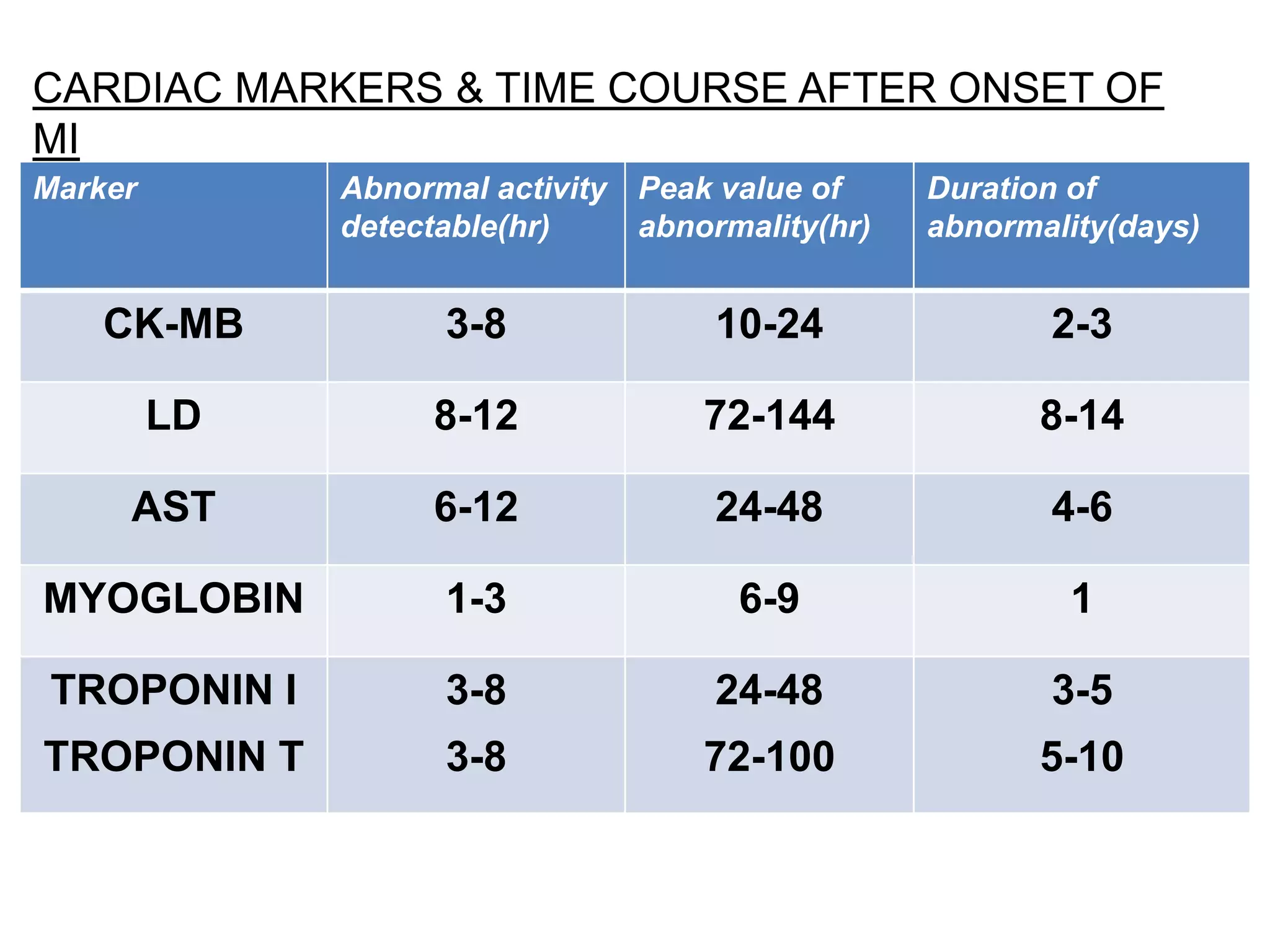
Troponin:
- Troponin I (cTnI). This kind of troponin is unique to heart muscle.
- Troponin T (cTnT).
Troponin T does exist in other types of muscle, but the amounts are
very limited. The Troponin T in your heart muscle also has a slightly
different structure, which doesn’t occur anywhere else in your body. Link
Newer, high-sensitivity tests can often detect even the tiny amounts of troponin in your blood that happen normally. In these cases, providers will repeat the test. If they see an increase in the troponin level on
the repeat test, that’s an indication of heart muscle damage. Link
The test should be repeated 12 hours after the onset of the peak symptoms. If any patient has a hs-cTnT level >14ng/L they should have a second sample sent for hs-cTnT testing six hours later. Link
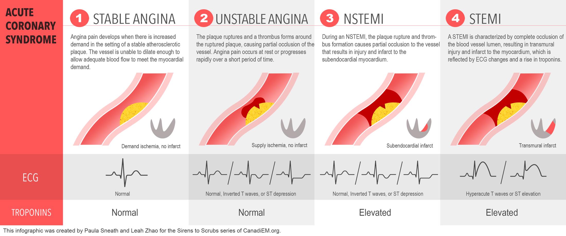
- Angina x MI
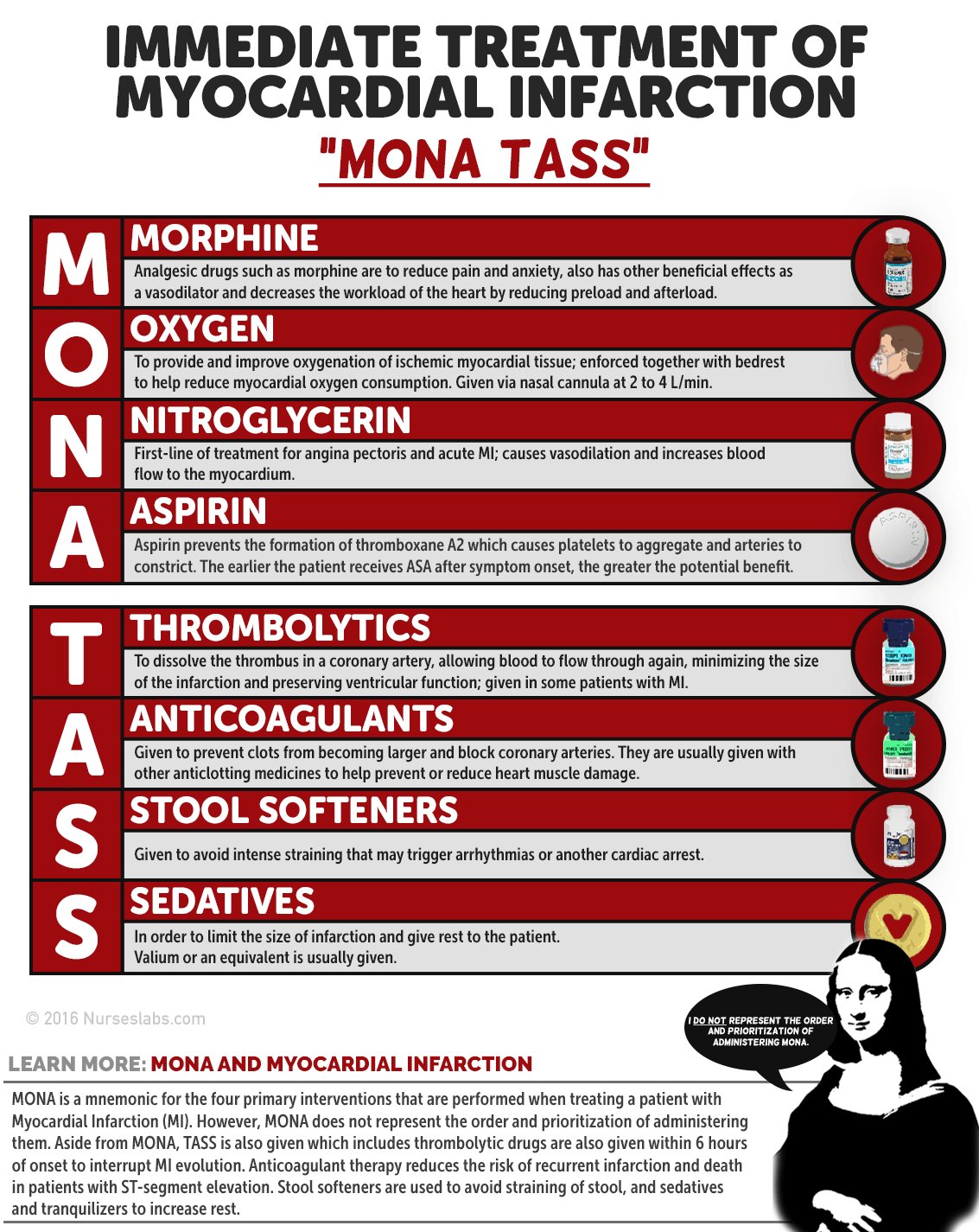
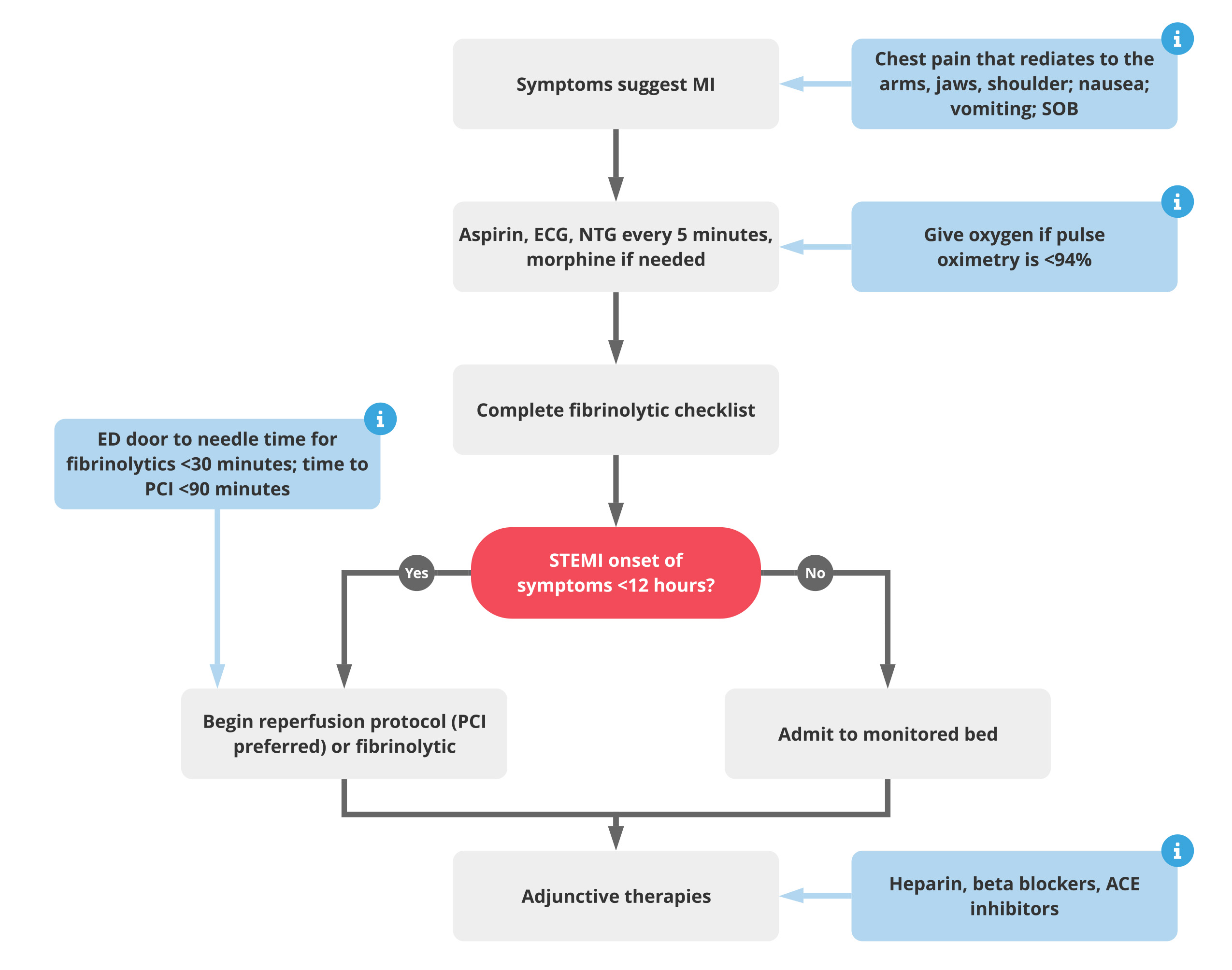
- ACS Rx